


Overexpression of ZNF169 promotes the growth and proliferation of colorectal cancer cells via the upregulation of ANKZF1
- Authors:
- Published online on: April 23, 2024 https://doi.org/10.3892/or.2024.8741
- Article Number: 82
-
Copyright: © Zhang et al. This is an open access article distributed under the terms of Creative Commons Attribution License.
Abstract
Introduction
Colorectal cancer (CRC) is the third most frequent malignancy and the second leading cause of cancer-associated mortality worldwide (1). An early diagnosis is crucial for the survival of patients with CRC. The 5-year survival rate of patients with early-stage CRC can be as high as 90%, whereas this rate decreases to only ~12% for patients with distant metastases (2). Colonoscopy is a well-established method for the diagnosis of CRC; however, its invasive nature limits its use for certain patients (3). Therefore, more accurate and less invasive screening methods are required for the detection of CRC. Over the past few decades, alongside the rapid development of tumor biotechnology, including genomics (4), transcriptomics (5), proteomics (6), metabonomics (7) and non-coding RNA omics (8), intensive studies have been conducted on the mechanisms of CRC tumorigenesis. This has led to the discovery of novel biomarkers for the diagnosis of CRC and drug targets for treatment (9,10). CRC is a heterogeneous disease (11), and factors such as tumor cell differentiation and proliferation markedly affect disease development, including metastases, disease outcomes and the response to anti-tumor drugs (12). However, the mechanisms through which these tumor heterogeneity factors are associated with CRC malignancy are not yet well understood (13); therefore, identifying novel drivers for CRC progression may shed light on the tumorigenesis of CRC, and may provide targets for diagnosis and therapy.
Zinc finger proteins (ZNFs), which contain a wide variety of zinc finger domains, are one of the largest protein families in eukaryotes (14). ZNFs are able to interact with DNA, RNA, poly-ADP-ribose (PAR) and other proteins, participating in multiple cellular processes, such as transcriptional regulation, ubiquitin-mediated protein degradation, signal transduction, DNA repair and cell migration (15). Increasing evidence has demonstrated that ZNFs play essential roles in the progression of various types of cancer, including lung, esophageal, colorectal, nasopharyngeal, thyroid, gastric, breast, ovarian, cervical, prostate and gallbladder cancer (16–18). ZNF169 is a C2H2-type zinc finger with Kruppel associated box domain (19). The ZNF169 gene is located at the D9S196-D9S280 interval on chromosome 9q22.3 (20). The expression of the ZNF169 gene has been found in multiple organs and tissues, including the alimentary tract (21). Although the physiological and pathological functions of ZNF169 are not yet completely clear, a previous study reported that its dysregulation may be involved in the progression of skin cancer (22). Via bioinformatic analysis of The Cancer Genome Atlas (TCGA) database, the clinical relevance and correlations of ZNF169 with genes in patients with CRC were revealed. However, the precise function and mechanisms of action of ZNF169 in CRC remain to be elucidated. Therefore, it is meaningful to conduct experiments to elucidate the significance of ZNF169 during the development of CRC.
Ankyrin repeat and zinc-finger domain-containing 1 (ANKZF1; also known as ZNF744), another ZNF, is a peptidyl-tRNA hydrolase and a co-factor of Cdc48 (yeast homolog to p97). The present study analyzed the genes positively correlating with ZNF169 in CRC tissues based on TCGA database. ANKZF1 was revealed to be one of the 10 of the most significantly positive genes, Unlike ZNF169, ANKZF1 is located in the cytoplasm, and is a component of the ribosome quality control complex that binds to ribosomes and releases nascent chains from tRNA, resulting in degradation and subsequent protein synthesis (23). ANKZF1 plays a role in the regulation of cellular functions, including cell cycle, apoptosis, autophagy and DNA damage repair (24). It was recently reported that ANKZF1 expression was associated with the prognosis of patients with colon cancer (25,26), and that it may be a promoting factor in the development of infantile-onset inflammatory bowel disease (IBD) (27). However, the upstream regulator of ANKZF1 in CRC remains unclear.
The present study aimed to investigate the clinical significance, cellular function and underlying mechanisms of ZNF169 in CRC cell growth and proliferation based on the analysis of TCGA database, the immunohistochemical staining (IHC staining) of CRC patient tissues and gain-of-function/loss-of-function experiments on CRC cell lines.
Materials and methods
IHC staining of ZNF169 and ANKZF1
The IHC staining of a human CRC tissue microarray was conducted by Servicebio Biotechnology Co., Ltd. The paired CRC tissues and adjacent normal tissues (at a distance of 5 cm from the tumor margin) were collected from patients with CRC undergoing surgical treatment between September, 2022 and March, 2023. The inclusion criteria were patients with primary pathologically diagnosed CRC. The exclusion criteria were patients with CRC had been treated with hormonal therapy or chemoradiotherapy, or patients with chronic inflammatory disease, severe cerebro-cardiovascular disease, respiratory disease, liver or renal disease, or female patients who were pregnant. The tissue sections (5-µm-thick) were subjected to IHC staining for ZNF169 and ANKZF1. Briefly, the sections were deparaffinized in xylene and hydrated in a graded alcohol series. Antigen retrieval and endogenous peroxidase blocking were performed using citrate buffer (pH 6) (cat no. G1219, Wuhan Servicebio Technology Co., Ltd.) and 3% hydrogen peroxide (cat no. 88597, MilliporeSigma), respectively. The slides were incubated with 10% goat serum (BIOSS) for 1 h and with rabbit polyclonal ZNF169 antibody (1:1,000 dilution; cat no. B-10117, Weibo Biotechnology Co., Ltd.) and rabbit polyclonal ANKZF1 primary antibody (1:500 dilution; cat no. 20446-1-AP, Proteintech Group, Inc.) at 4°C overnight. The antigen-antibody complex was detected with a biotinylated goat anti-rabbit antibody (1:2,500 dilution; cat no. BA1003, Boster Biological Technology Co., Ltd.) conjugated with streptavidin-horseradish peroxidase was used to detect the antigen-antibody complex. Visualization was conducted using nickel-enhanced 3,3-diaminobenzidine tetrahydrochloride (Beijing Solarbio Science & Technology Co., Ltd.). The tissue sections were then counterstained with hematoxylin (cat no. G1004, Wuhan Servicebio Technology Co., Ltd.) for 3–5 min at room temperature. For the mean density analysis of IHC staining, three or more fields (magnification, ×200) were randomly selected from each section for imaging. The staining density value is the relative expression level of the proteins. The high or low expression of ZNF169 and ANKZF1 was set during the density analysis. The present study was approved by the Ethics Committee of Beijing Rehabilitation Hospital of Capital Medical University (Beijing, China). A form of written informed consent was obtained from each participant in the present study.
Analysis of ZNF169 and ANKZF1 based on TCGA
The transcript level of ZNF169 and ANKZF1, the Spearman's correlation analysis of the correlation between ZNF169 and other genes, as well as the prognostic significance of ZNF169 and ANKZF1 in patients with CRC were analyzed based on TCGA database (http://cancergenome.nih.gov).
Cells and cell culture
The 293T cells (cat no. CRL-3216) was purchased from the American Type Culture Collection. The normal human colorectal cell line NCM460 (cat no. CL0393) was purchased from FENGHUIBIO (www.fenghbio.cn). The CRC cell lines, RKO (cat no. CL-0196), HCT-116 (cat no. CL-0096), HT-29 (cat no. CL-0118), SW620 (cat no. CL-0225) and HCT-8 (cat no. CL-0098), were purchased from Procell Life Science & Technology Co. Ltd. STR profiling was used to confirm the authentication of the HT-29 cell line. All the cells were cultured in RPMI-1640 complete medium (Gibco; Thermo Fisher Scientific, Inc.), containing 10% fetal bovine serum (Gibco; Thermo Fisher Scientific, Inc.) and 1% penicillin/streptomycin solution (Corning, Inc.). The cells were maintained in 6-cm plates at 37°C in an incubator with 5% CO2.
Knockdown and overexpression of ZNF169 and ANKZF1
For the knockdown assay, the HCT-116, RKO or HT-29 CRC cells were transfected with small interfering RNAs (siRNAs) against ZNF169, ANKZF1 or negative control siRNA using RNAiMAX regent (Invitrogen; Thermo Fisher Scientific, Inc.). A total of 3 µl of 20 µM siRNAs and 4 µl RNAiMAX were added to 400 µl Opti-MEM (cat no. 31985070, Gibco; Thermo Fisher Scientific, Inc.) and were incubated at room temperature for 15 min. The mixture was then added to 1.6 ml culture medium. After 48 h, the cells were examined using reverse transcription-quantitative PCR (RT-qPCR), western blot analysis, cellular function assays and other experiments. The siRNAs were purchased from HIPPOBIO company (http://www.hippobiotec.com/). The siRNA sequences were as follows: siCtrl, 5′-UUCUCCGAACGUGUCACGU-3′; siZNF169, 5′-GAAGCUCCAAGAUGCUCUAGU-3′; siANKZF1, 5′-GGUGCUAUAUUUCAAGGAAGA-3′.
For the overexpression assay, the coding sequences of ZNF169 or ANKZF1 were synthesized and cloned into the pCDH overexpression plasmids by HIPPOBIO company (http://www.hippobiotec.com/). Subsequently, lentiviruses were produced by transfecting the 293T cells using VigoFect (cat no. Vigorous Biotechnology, http://www.vigorousbiol.com/index.htm) with pCDH-Ctrl, pCDH-ZNF169, or pCDH-ANKZF1 vectors, alongside the packaging plasmids, including PSPAX2 and PDM2G. After 48 h, the virus supernatants were harvested, filtered and concentrated. Subsequently, the lentiviruses were added to the medium, supplied with polybrene, to infect the HT-29 or HCT-116 cells for 48 h. Finally, the cell lines with a stable overexpression were constructed by treating the cells with puromycin (cat no. HB-PU-1000, HANBIO, http://www.hanbio.net/) for 2 weeks.
RT-qPCR
Total RNA was extracted from the HCT-116, RKO or HT-29 cells using TRIzol® reagent (Invitrogen; Thermo Fisher Scientific, Inc.), according to the manufacturer's instructions. ReverTra Ace™ qPCR RT Master Mix with gDNA Remover kit (cat no. FSQ-301, Toyobo Life Science) was then applied to reverse transcribe the RNA into cDNA, according to the manufacturer's instructions. Subsequently, the quantification of cDNA was performed using qPCR with SYBR master mixture (Takara Bio, Inc.) on a Bio-Rad system (Bio-Rad Laboratories, Inc.). The PCR thermocycling conditions were as follows: i) Holding stage, 95°C for 3 min; ii) cycling stage, 95°C for 15 sec; iii) Cycling stage, 60°C for 30 sec; iv) cycling stage, 72°C for 30 sec; cycling stage, 2–4; 40 cycles. The qPCR primer sequences were as follows: ANKZF1 forward, 5′-CGGTTTAACCTAAAGCAACGTCT-3′ and reverse, 5′-CCGATCCAGTGTCTGCAAGT-3′; β-actin forward, 5′-CATGTACGTTGCTATCCAGGC-3′ and reverse, 5′-CTCCTTAATGTCACGCACGAT-3′. The quantification of qPCR data was performed according the 2−ΔΔCq method (28).
Western blot analysis
Total protein was extracted from the NCM460, RKO, HCT-116, SW620, HT-29 and HCT-8 cells using RIPA buffer (Beyotime Institute of Biotechnology). The protein concentration was measured using the BCA kit (Thermo Fisher Scientific, Inc.). A total of 40–70 µg proteins were then separated by SDS-PAGE on 10 or 12% gels, followed by transfer onto PVDF membranes. The PVDF membranes were blocked with 5% non-fat milk and incubated with primary antibodies at 4°C overnight. After washing with PBS (Wuhan Servicebio Technology Co., Ltd.)-Tween (Beyotime Institute of Biotechnology), the membranes were incubated with secondary antibodies for 2 h at room temperature. The protein signal was detected using the ECL-Plus kit (cat no. RPN2232, Cytiva). The antibody against ZNF169 (1:1,000; cat. no. ab225924) was purchased from Abcam, and the antibodies against ANKZF1 (1:800; cat. no. 20447-1-AP) and GAPDH (1:10,000; 10494-1-AP) were obtained from Proteintech Group, Inc. The mouse anti-rabbit IgG-HRP secondary antibody (1:8,000; cat. no. sc-2357) was from Santa Cruz Biotechnology, Inc.
Cell Counting Kit-8 (CCK-8)
The proliferation of the CRC cells was determined using a CCK-8 kit. Briefly, a total of 3,000 HCT-116, RKO and HT-29 cells were seeded into 96-well plates, which contained 200 µl culture medium. After 8 h, 20 µl CCK-8 regent (cat no. C0039, Beyotime Institute of Biotechnology) were added to each well and incubated at 37°C for 3 h. The OD value was then measured at 450 nm, which represented the relative cell viability of 0 h. At the time points of 24, 48, 72 and 96 h, the cell viability was sequentially determined to compare to the OD450 value at 0 h.
Colony formation assay
Following ZNF169 knockdown, 2,000 HCT-116 and RKO cells from the siCtrl and siZNF169 groups were seeded into six-well plates, and the cells in completed culture medium were maintained for 8 days in a cell incubator at 37°C. Following the overexpression of ZNF169, 500 HT-29 cells from the Ctrl and ZNF169 overexpression groups were cultured in six-well plates for 10 days. After colonies were formed, they were washed twice with PBS (Wuhan Servicebio Technology Co., Ltd.). Subsequently, methanol was used to fix the colonies for 30 min at room temperature and 0.1% crystal violet (Beyotime Institute of Biotechnology) was used to stain them for 30 min at room temperature. Finally, the images of the colonies were captured using a camera (EOS700D, Canon, Inc.).
Measurement of caspase-3/7 activity
Caspase-3/caspase-7 activity was detected using the Caspase-Glo reagent (Promega Corporation). Briefly, 10,000 suspended HCT-116, RKO or HT-29 cells were seeded into 96-well plates, to which 100 µl Caspase-Glo was added. After 90 min, the caspase-3/caspase-7 activity was detected using a microplate reader (M2009PR, Tecan infinite, Tecan Group, Ltd.).
EdU staining
To assess DNA synthesis, the HCT-116, RKO or HT-29 CRC cells were subjected to EdU staining using the BeyoClick™ EdU Cell Proliferation kit with Alexa Fluor 555 (Beyotime Institute of Biotechnology), according to the manufacturer's instructions. A total of 5×10⁵ HCT-116 and RKO cells transfected with a negative control siRNA or a siRNA against ZNF169, or a total of 4×10⁵ HT-29 cells infected with an empty control or ZNF169 overexpression lentivirus, were seeded onto coverslips in six-well plates. After 24 h, the cells were incubated with EdU reagent at 37°C for 4–6 h. The cells on the coverslips were then washed with PBS, fixed with 4% paraformaldehyde (Wuhan Servicebio Technology Co., Ltd.) for 15 min at room temperature, treated with 0.5% Triton X-100 (Beyotime Institute of Biotechnology) for 15 min at room temperature, and finally stained with Click Additive Solution (Beyotime Institute of Biotechnology) for 30 min at room temperature and DAPI (Beyotime Institute of Biotechnology) for 15 min at room temperature in the dark. Images of the EdU-positive cells were captured under a microscope and quantified by Photoshop.
Dual luciferase reporter assay
The promoter of ANKZF1 was cloned into the pGL3 basic vector (Promega Corporation). The HCT-116 and HT-29 cells were seeded in 24-well plates and co-transfected with the aforementioned luciferase vectors and the expression vectors (knockdown or overexpression vector) using VigoFect for 48 h in a cell incubator at 37°C. The Renilla luciferase vector, pCMV-RL-TK (Promega Corporation), was used as an internal control to determine the transfection efficiency. At 48 h following transfection, the dual luciferase activity was checked by using the Dual-Luciferase® Reporter Assay System (E1910, Promega Corporation), according to the manufacturer's protocols.
Chromatin immunoprecipitation (ChIP)-qPCR
A ChIP assay was performed to determine whether ZNF169 binds to the promoter of the ANKZF1 gene in the control and ZNF169-overexpressing cells. The ChIP-qPCR assay was performed using the SimpleChIP enzymatic chromatin IP kit (cat no. 26156, Thermo Fisher Scientific, Inc.), according to the manufacturer's protocols. The purified DNA was then subjected to qPCR analysis. The qPCR primers were designed to amplify the promoter sequence (2,000 bp upstream of the ANKZF1 transcript) of the ANKZF1 gene. The qPCR primer sequences were as follows: Forward, 5′-CTCCATGTTCTTCCATCAACT-3′ and reverse 5′-CTCCTTATTGGCAGGTGGTC-3′.
Statistical analysis
GraphPad Prism 8.0 software (Dotmatics) was used for statistical analyses. The results are presented as the mean ± standard error of the mean (meean ± SEM). Adobe Illustrator 2021 (Adobe Inc.) was utilized to create the figures. The Chi-squared test (χ2 value) was performed to assess the relevance of ZNF169 expression in CRC and normal samples. An unpaired Student's t-test and one-way ANOVA followed by Tukey's post hoc test were applied to compare the differences between two groups and among more than two groups, respectively. P<0.05 was considered to indicate a statistically significant difference.
Results
ZNF169 expression is upregulated in patients with CRC
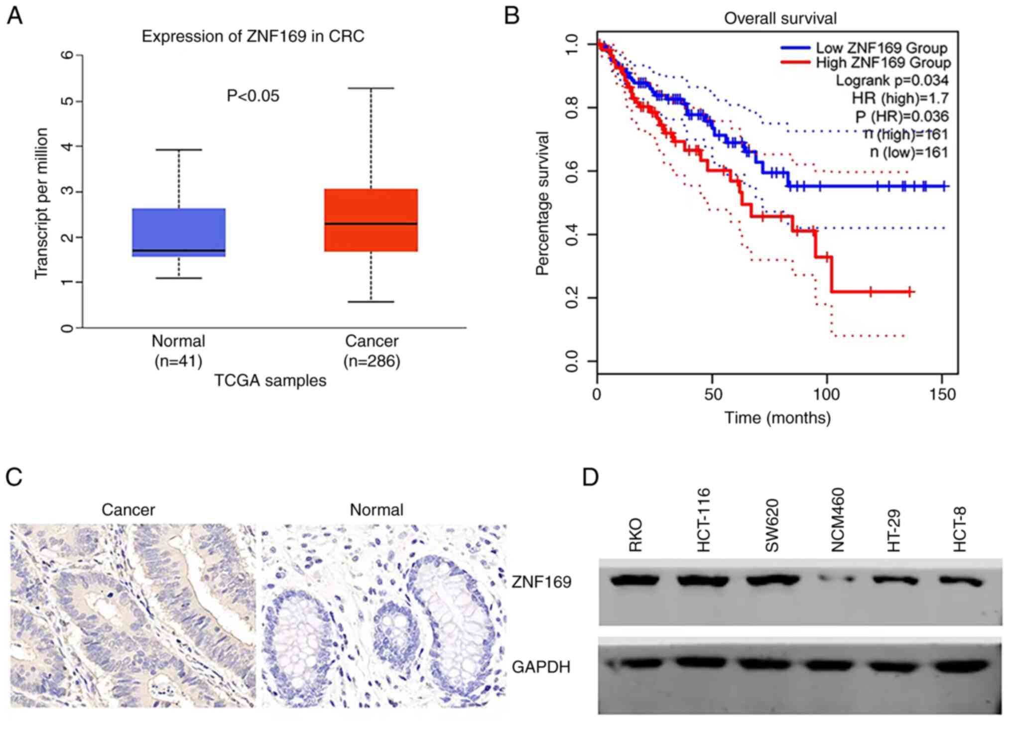
To examine the clinical relevance of ZNF169 in CRC, the present study first collected data from TCGA and analyzed the expression of ZNF169 in CRC and normal tissues. It was found that ZNF169 expression was upregulated in CRC tissues compared with normal tissues (Fig. 1A). GEPIA is a newly developed interactive web server which uses the log-rank test (the Mantel-Cox test) for survival analysis (29). The patients with CRC were subsequently divided into the ZNF169 high and low expression groups, and their survival rates were analyzed from GEPIA. The patients in the high ZNF169 expression group had a shorter survival time (Fig. 1B), suggesting that ZNF169 functions as a negative prognostic biomarker for patients with CRC. In order to validate the protein expression of ZNF169, a tissue microarray containing cancer and normal tissues from patients with CRC was subjected to the IHC staining of ZNF169. The results indicated that the protein expression of ZNF169 was higher in CRC tissues than in normal tissues (Fig. 1C and Table I). It was also found that ZNF169 was highly expressed in CRC cell lines, including RKO, HT-29 and HCT-116, as compared with the NCM460 normal cell line (Fig. 1D).
ZNF169 promotes the proliferation and growth of CRC cells
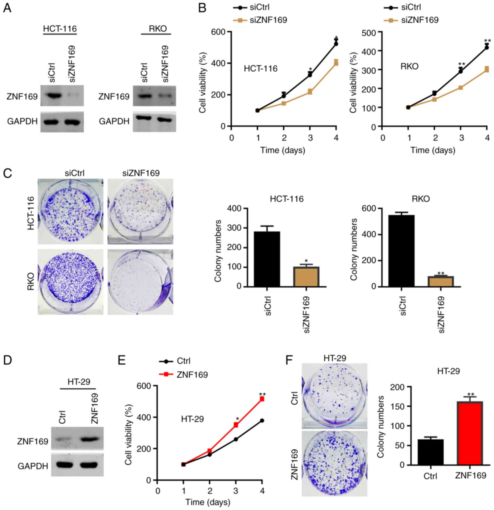
The present study then explored the cellular function of ZNF169 in CRC cells. As the HCT-116 and RKO cells had a relatively higher expression of ZNF169, these cells were transfected with a negative control siRNAs and with siRNAs against and ZNF169. The results of western blot analysis revealed that ZNF169 was efficiently silenced in the siZNF169 cells (Fig. 2A). The results of the CCK-8 assay demonstrated that ZNF169 knockdown suppressed the proliferation of the HCT-116 and RKO cells (Fig. 2B). Consistently, the colony formation capacity of the HCT-116 and RKO cells was also suppressed following the knockdown of ZNF169 (Fig. 2C). Subsequently, the HT-29 cells were transfected with an empty control or ZNF169 overexpression lentivirus, and the results of western blot analysis validated the overexpression efficacy (Fig. 2D). The results of CCK-8 and colony formation assays demonstrated that the ectopic overexpression of ZNF169 significantly promoted the malignant growth of the HT-29 cells (Fig. 2E and F). These results suggested that ZNF169 may be critical for the growth and proliferation of CRC cells.
ZNF169 potentiates EdU staining and inhibits caspase activity in CRC cells
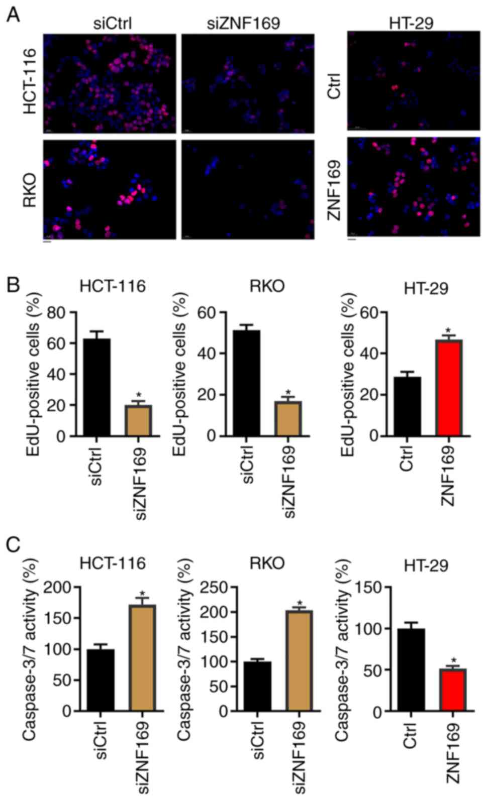
To examine the mechanisms through which ZNF169 contributes to the proliferation of CRC cells, the CRC cells in which ZNF169 was knocked down or overexpressed were subjected to EdU staining, which predicts DNA synthesis. It was found that ZNF169 knockdown significantly suppressed the proportion of EdU-positive cells in both cell lines (Fig. 3A and B). By contrast, the ectopic overexpression of ZNF169 enhanced the proportion of EdU-positive HT-29 cells (Fig. 3A and B). The present study then analyzed the activity of caspase-3/7, which can be used to predict apoptosis, following the knockdown or overexpression of ZNF169 in the cells. As shown in Fig. 3C, ZNF169 knockdown enhanced caspase-3/7 activity in the HCT-116 and RKO cells. Conversely, ZNF169 overexpression suppressed caspase-3/7 activity in the HT-29 cells. These results indicated that ZNF169 promotes DNA synthesis and suppresses the apoptosis of CRC cells.
ZNF169 transcriptionally activates ANKZF1 in CRC cells
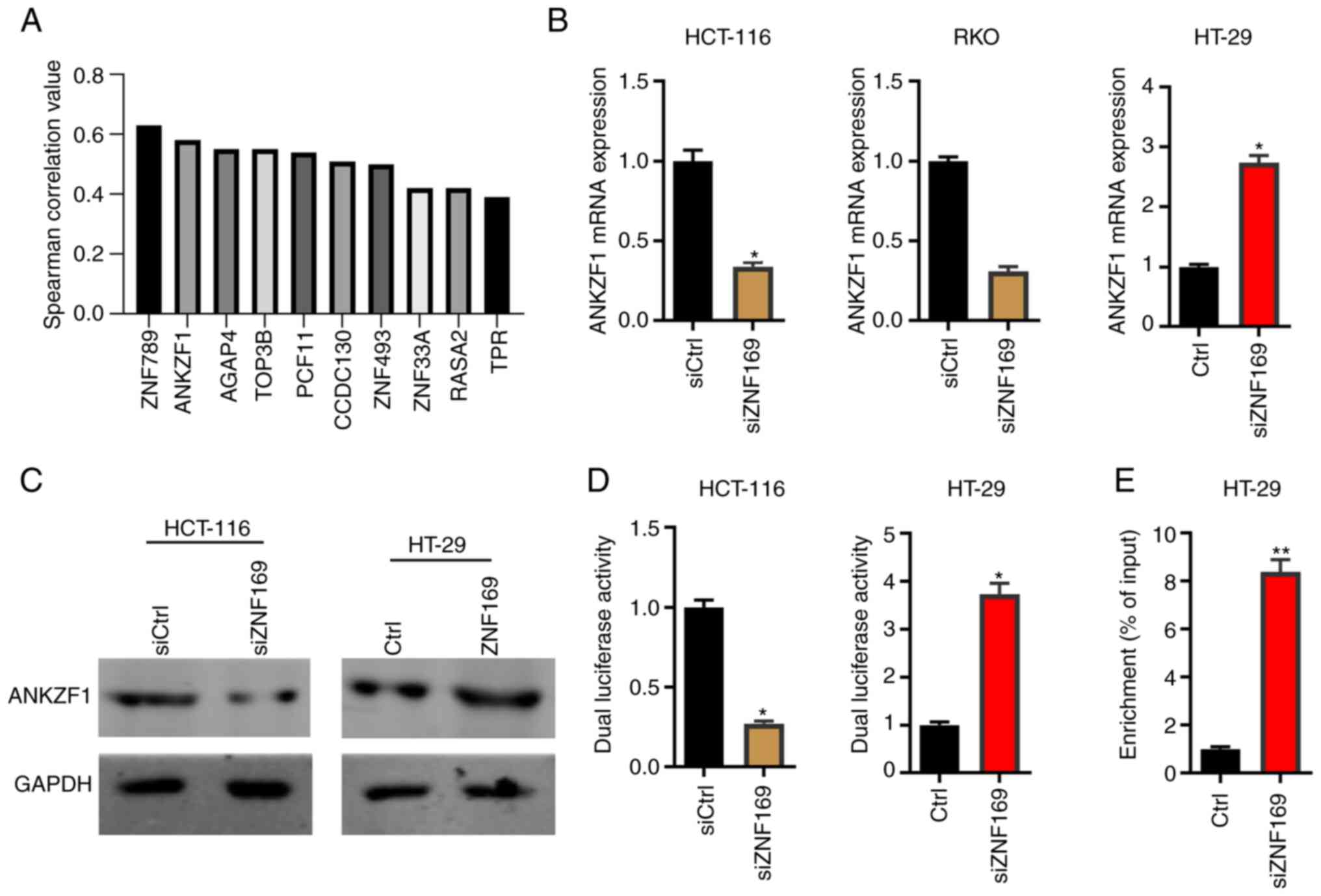
Since ZNF169 serves as a transcription factor in eukaryotes, there must be downstream effectors that participate in the ZNF169-induced promotion of CRC cell proliferation. Therefore, the present study then analyzed the genes positively correlated with ZNF169 in CRC tissues based on TCGA database. A total of 10 of the most significantly positively correlated genes, including ZNF789 and ANKZF1, are illustrated in Fig. 4A. Since the overexpression of ANKZF1 has been reported to be associated with the poor prognosis of patients with CRC (25,26), the present study aimed to explore whether ZNF169 regulated ANKZF1 in the present study, and we aim to explore the significance of other genes positively correlated with ZNF169, such as ZNF789 and AGAP4, in the subsequent studies.. Based on the results of RT-qPCR and western blot analysis, it was found that ZNF169 knockdown markedly suppressed the mRNA and protein expression levels of ANKZF1 in the HCT-116 and RKO cells (Fig. 4B and C). By contrast, the overexpression of ZNF169 potentiated the expression of ANKZF1 in the HT-29 cells (Fig. 4B and C). In order to examine whether ZNF169 regulates the transcriptional activity of the ANKZF1 gene, the promoter sequence of the ANKZF1 gene was cloned into a pGL3.basic vector and a dual luciferase activity assay was performed. It was demonstrated that ZNF169 knockdown inhibited the luciferase activity of the ANKZF1 promoter, whereas ZNF169 overexpression promoted it (Fig. 4D). In addition, a ChIP-qPCR assay was carried out to examine the interaction between the ZNF169 protein and the promoter sequence of the ANKZF1 gene. It was found that the promoter sequence of the ANKZF1 gene was highly enriched in the ANKZF1-Flag group of CRC cells (Fig. 4E). Collectively, ZNF169 may potentiate the transcriptional activity of the ANKZF1 gene by directly interacting with its promoter sequence.
ZNF169 contributes to the proliferation of CRC cells through upregulation of ANKZF1
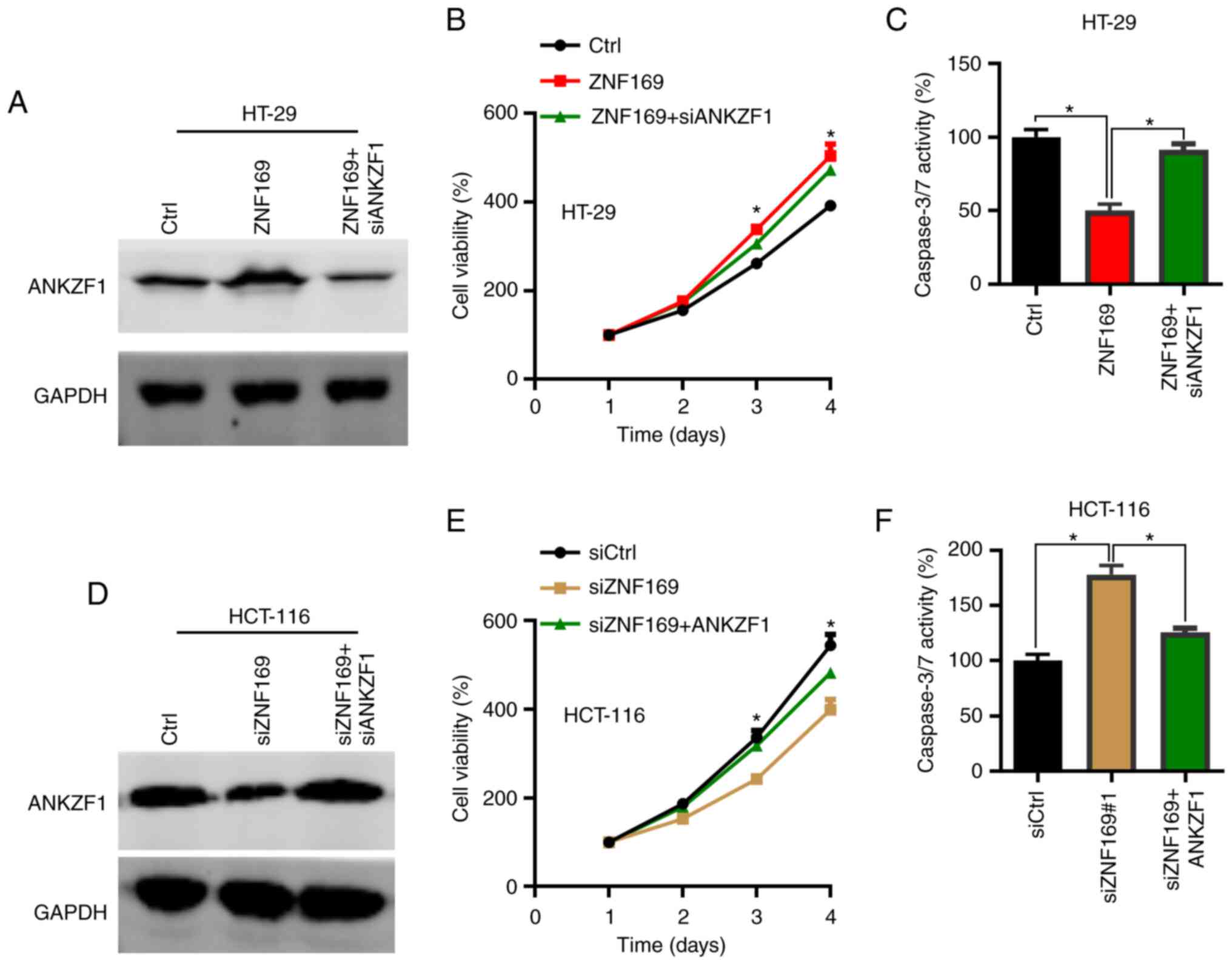
As ZNF169 was found to promote CRC cell proliferation and positively regulate the expression of ANKZF1, the present study then aimed to determine whether ANKZF1 participates in CRC growth. To this aim, overexpression and knockdown assays of ANKZF1 were performed in the CRC cells. The results of RT-qPCR revealed that ANKZF1 could be efficiently silenced by siANKZF1 when compared with siCtrl in the HT-29 cells (Fig. S1A). Thus, ANKZF1 was knocked down (Fig. S1B) in ZNF169-overexpressing CRC cells and the cells were subjected to CCK-8 and colony formation assays. The results of western blot analysis revealed that ANKZF1 was efficiently silenced after the ZNF169-overexpressing cells were transfected with siANKZF1 (Fig. 5A). The results of CCK-8 and colony formation assays demonstrated that ANKZF1 knockdown suppressed the growth and proliferation of the CRC cells in which ZNF169 was overexpressed (Fig. 5B and C). To validate the function of ANKZF1, ANKZF1 was then overexpressed in the HCT-116 cells in which ZNF169 was silenced. ANKZF1 overexpression lentivirus significantly increased the mRNA level of ANKZF1 in the HCT-116 cells when compared with the cells transfected with the Ctrl lentivirus (Fig. S1B). Based on the results of western blot analysis, ANKZF1 was found to be overexpressed in the HCT-116 cells in which ZNF169 was silenced (Fig. 5D). ANKZF1 overexpression restored the proliferation and growth of the HCT-116 cells in which ZNF169 was knocked down (Fig. 5E and F). These results suggested that ZNF169 may contribute to the growth and proliferation of CRC cells through the upregulation of ANKZF1.
ANKZF1 overexpression is associated with the poor prognosis of patients with CRC
Finally, the clinical relevance of ANKZF1 was investigated in patients with CRC. Based on TCGA database, ANKZF1 expression was upregulated in the cancer tissues of patients with CRC compared with normal tissues (Fig. 6A). Consistently, the results of IHC staining revealed that the protein expression of ANKZF1 was higher in CRC tissues compared with adjacent normal tissues (Fig. 6B). It was also demonstrated that there was a positive correlation between ZNF169 and ANKZF1 expression in the tissues of patients with CRC (Fig. 6C and Table II). Importantly, a high expression of ANKZF1 was associated with a poor prognosis of patients with CRC as compared with those with a low expression of ANKZF1 (Fig. 6D). Thus, ANKZF1 may be considered a negative prognostic biomarker for patients with CRC.
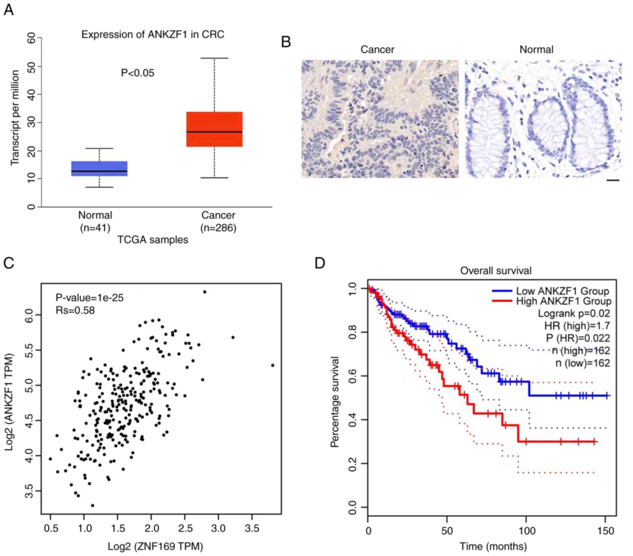
Table II.Results of Spearman's correlation analysis of the correlation between ZNF169 and ANKZF1 expression in 22 CRC tissues examined using immunohistochemistry. |
Discussion
Members of the ZNF family can function as either tumor suppressor genes or oncogenes (16). Recently, the importance of ZNFs in cancer onset and progression has been actively studied. The findings have demonstrated that ZNFs are involved in the regulation of the malignancy of cancer cells through a variety of mechanisms, including by functioning as transcription factors (15,30). The results from the present study demonstrated that ZNF169 functioned as an oncogene to promote the development of CRC by potentiating the expression of ANKZF1. To the best of our knowledge, this is the first study on the role of ZNF169 in tumorigenesis. ZNF169 is considered to function as a transcription factor, and may have functions in DNA-binding transcription activity, particularly in RNA polymerase II transcription regulatory region sequence-specific DNA-binding activity (31,32). RNA polymerase II transcribes all protein-coding genes in eukaryotic genomes (33); therefore, ZNF169 could indirectly affect various biological processes. ZNF169 has been reported to be relevant to obesity, malignant essential hypertension, speech-language disorder-1, Fanconi anemia and other human disorders (22,34,35). However, both the regulation of ZNF169 on target genes and the regulation of ZNF169 expression are not yet completely clear. Bhattacharya and Ghosh (36) demonstrated that herpes virus-associated ubiquitin specific protease is implicated in the regulation of transcription factors, including ZNF169. Huttlin et al (37) employed robust affinity purification-mass spectrometry methodology and demonstrated that ZNF169 interacts with dozens of other proteins; however, the precise results of these interactions need to be further investigated.
The data of the present study demonstrated that ZNF169 promoted DNA synthesis and reduced caspase-3 and −7 activity in CRC cell lines. This may be one of the mechanisms underlying the promoting effects of ZNF169 on CRC malignancy. These results further indicated the oncogenic role of ZNF169 in the progression of CRC. The significance of ZNFs has been reported in CRC development by other studies. Cheng et al (38) found that ZNF277 was overexpressed in human CRC samples. Xie et al (39) further confirmed that ZNF277 was uniquely expressed in early colorectal stem cells and undifferentiated transit-amplifying cells. In addition, it was reported that the overexpression of ZNF277 is critical for maintaining CRC growth (39). ZNF367 is another oncogene in CRC. The depletion of ZNF367 has been reported to blunt the proliferation and invasion of CRC cells via the inactivation of the YAP signaling pathway (40). In CRC, ZNF281 also plays a role in metastasis through the regulation of the epithelial-mesenchymal transition (EMT). During EMT, c-MYC induces the expression of ZNF281 in a SNAIL-dependent manner, while microRNA (miR)-34a decreases the post-transcriptional level of ZNF281. Notably, p53 can promote the expression of miR-34a (41). ZNF281, functioning as a regulator, has been shown to be associated with an increased migration/invasion and an enhanced β-catenin activity (42). There are numerous proteins in the ZNF family, and their functions have a wide range of molecular effects on several cellular processes (15). It has been reported that ZNFs can affect cancer malignancy via various mechanisms (16). A single ZNF can function as a suppressor gene in certain types of tumors, whereas it may function as an oncogene in others. ZBP89 (also referred to as ZNF148) is C2H2-type transcription factor. On the one hand, ZBP89 exerts its oncogenic function by regulating matrix metallopeptidase 3, and inhibiting ornithine decarboxylase and vimentin, in breast cancer, melanoma and gastric cancer (43–45). These three molecules are involved in tumor development, migration, invasion and metastasis. On the other hand, ZBP89 may function as a tumor suppressor gene by inhibiting cell proliferation and inducing apoptosis by regulating the β-catenin pathway in CRC (46,47).
In the present study, it was found that ZNF789 and ANKZF1 were the top two genes which positively correlated with the expression of ZNF169 in the tissues of patients with CRC. To the best of our knowledge, the involvement of ZNF789 has not been reported in cancer. The present study focused on ANKZF1 and identified ANKZF1 as a downstream effector in the ZNF169-induced promotion of CRC cell proliferation. ZNF169 positively regulated the expression of ANKZF1 by directly interacting with the promoter of the ANKZF1 gene, and ANKZF1 contributed to the accelerated proliferation of CRC cells triggered by ZNF169. Apart from the regulation of ANKZF1 by ZNF169, there may be different pathways involved in the regulation of ANKZF1 expression, such as the IFN receptor family (48). Lai and Park (49) demonstrated that MET, also known as hepatocyte growth factor receptor, was involved in the regulation of the transcription of the ANKZF1 gene in gastric cancer cells, as the expression of the ANKZF1 gene was shown to be increased when the cells were treated with a MET molecule inhibitor. Furthermore, the activity of peroxisome proliferator-activated receptor could inhibit the transcription of ANKZF1 in monocyte-derived dendritic cells (50). It has been reported that the increased expression of ANKZF1 may predict the malignant progression and poor survival of patients with CRC (25,51). by contrast, the knockdown of ANKZF1 suppresses the growth and invasion of CRC cells (52). These results suggest that ANKZF1 may function as an oncogenic protein in CRC. van Haaften-Visser et al (27) demonstrated that ANKZF1 mutations lad to a reduction in mitochondrial integrity and respiration, which may be a pathological factor in the pathogenesis of infantile-onset IBD.
A number of issues remain unclear, such as the mechanisms underlying the effects of the upregulation of ZNF169 expression in patients with CRC and the additional mechanisms upregulating ANKZF1 expression in CRC. These questions need to be addressed in future studies. DNA methylation of gene promoters and copy number variants of genes are two key events contributing to the dysregulation of genes; however, it remains to be determined whether these events play a role in the expression of ZNF169 and/or ANKZF1 in CRC. It may be worthwhile to explore the role of the upstream or downstream factors in the regulation of the ZNF169/ANKZF1 axis.
In conclusion, the present study demonstrated that the expression of ZNF169 was markedly increased in CRC, and high expression pf ZNF169 was associated with the poor overall survival of patients with CRC. Furthermore, ZNF169 could positively regulate its downstream factor, ANKZF1, which in turn accelerated CRC cell progression. The present study demonstrated the biological function of ZNF169 in CRC, which may provide novel insight into the diagnosis and treatment of CRC.
Supplementary Material
Supporting Data
Acknowledgements
Not applicable.
Funding
The present study was supported by the Scientific Research Fund of Beijing Rehabilitation Hospital, Capital Medical University (2019-043, 2019R-001 2020-056 and 2022-057), and the Scientific Research Fund of Beijing Anorectal Society (2020ABCP002).
Availability of data and materials
The datasets used and/or analyzed during the current study are available from the corresponding author on reasonable request.
Authors' contributions
QG, CK and HS designed the study. JZ, YW, SH, MX, MZ, JW, JS and XC performed the majority of the experiments. QG, CK and JZ analyzed the data. DD, YW, JW, JS and XC provided assistance for the experiments and data analysis. QG, CK HS and JZ wrote the draft of the manuscript. QG revised the manuscript. QG, CK and JZ confirm the authenticity of all the raw data. All authors have read and approved the final manuscript.
Ethics approval and consent to participate
The present study is approved by the Ethics Committee of the Beijing Rehabilitation Hospital of Capital Medical University (Beijing, China) (no. 2022bkkyLW003). A written form of informed consent was obtained from each participant in the present study.
Patient consent for publication
Not applicable.
Competing interests
The authors declare that they have no competing interests.
Glossary
Abbreviations
Abbreviations:
|
ANKZF1 |
ankyrin repeat and zinc-finger domain-containing 1 |
|
CCK-8 |
Cell Counting Kit-8 |
|
CRC |
colorectal cancer |
|
Chip-qPCR |
chromatin immunoprecipitation (Chip)-qPCR |
|
HIF-1α |
hypoxia-inducible factor 1α |
|
IHC staining |
immunohistochemical staining |
|
RT-qPCR |
reverse transcription-quantitative PCR |
|
TCGA |
The Cancer Genome Atlas |
|
ZNF169 |
zinc finger protein 169 |
References
|
Sung H, Ferlay J, Siegel RL, Laversanne M, Soerjomataram I, Jemal A and Bray F: Global cancer statistics 2020: GLOBOCAN estimates of incidence and mortality worldwide for 36 cancers in 185 countries. CA Cancer J Clin. 71:209–249. 2021. View Article : Google Scholar : PubMed/NCBI | |
|
Wu X, Lin H and Li S: Prognoses of different pathological subtypes of colorectal cancer at different stages: A population-based retrospective cohort study. BMC Gastroenterol. 19:1642019. View Article : Google Scholar : PubMed/NCBI | |
|
Kanth P and Inadomi JM: Screening and prevention of colorectal cancer. BMJ. 374:n18552021. View Article : Google Scholar : PubMed/NCBI | |
|
de Assis J, Coutinho L, Oyeyemi IT, Oyeyemi OT and Grenfell RFEQ: Diagnostic and therapeutic biomarkers in colorectal cancer: A review. Am J Cancer Res. 12:661–680. 2022.PubMed/NCBI | |
|
Sardo E, Napolitano S, Della Corte CM, Ciardiello D, Raucci A, Arrichiello G, Troiani T, Ciardiello F, Martinelli E and Martini G: Multi-Omic approaches in colorectal cancer beyond genomic data. J Pers Med. 12:1282022. View Article : Google Scholar : PubMed/NCBI | |
|
Zhu G, Jin L, Sun W, Wang S and Liu N: Proteomics of post-translational modifications in colorectal cancer: Discovery of new biomarkers. Biochim Biophys Acta Rev Cancer. 1877:1887352022. View Article : Google Scholar : PubMed/NCBI | |
|
Salita T, Rustam Y, Mouradov D, Sieber OM and Reid GE: Reprogrammed lipid metabolism and the Lipid-Associated hallmarks of colorectal cancer. Cancers (Basel). 14:37142022. View Article : Google Scholar : PubMed/NCBI | |
|
Yang M, Yang H, Ji L, Hu X, Tian G, Wang B and Yang J: A multi-omics machine learning framework in predicting the survival of colorectal cancer patients. Comput Biol Med. 146:1055162022. View Article : Google Scholar : PubMed/NCBI | |
|
Dekker E, Tanis PJ, Vleugels JLA, Kasi PM and Wallace MB: Colorectal cancer. Lancet. 394:1467–1480. 2019. View Article : Google Scholar : PubMed/NCBI | |
|
Andrei P, Battuello P, Grasso G, Rovera E, Tesio N and Bardelli A: Integrated approaches for precision oncology in colorectal cancer: The more you know, the better. Semin Cancer Biol. 84:199–213. 2022. View Article : Google Scholar : PubMed/NCBI | |
|
Ashktorab H and Brim H: Colorectal cancer subtyping. Nat Rev Cancer. 22:68–69. 2022. View Article : Google Scholar : PubMed/NCBI | |
|
Li J, Ma X, Chakravarti D, Shalapour S and DePinho RA: Genetic and biological hallmarks of colorectal cancer. Genes Dev. 35:787–820. 2021. View Article : Google Scholar : PubMed/NCBI | |
|
Sagaert X, Vanstapel A and Verbeek S: Tumor heterogeneity in colorectal cancer: What do we know so far? Pathobiology. 85:72–84. 2018. View Article : Google Scholar : PubMed/NCBI | |
|
Bu S, Lv Y, Liu Y, Qiao S and Wang H: Zinc finger proteins in neuro-related diseases progression. Front Neurosci. 15:7605672021. View Article : Google Scholar : PubMed/NCBI | |
|
Cassandri M, Smirnov A, Novelli F, Pitolli C, Agostini M, Malewicz M, Melino G and Raschellà G: Zinc-finger proteins in health and disease. Cell Death Discov. 3:170712017. View Article : Google Scholar : PubMed/NCBI | |
|
Jen J and Wang YC: Zinc finger proteins in cancer progression. J Biomed Sci. 23:532016. View Article : Google Scholar : PubMed/NCBI | |
|
Ecco G, Imbeault M and Trono D: KRAB zinc finger proteins. Development. 144:2719–2729. 2017. View Article : Google Scholar : PubMed/NCBI | |
|
Li X, Han M, Zhang H, Liu F, Pan Y, Zhu J, Liao Z, Chen X and Zhang B: Structures and biological functions of zinc finger proteins and their roles in hepatocellular carcinoma. Biomark Res. 10:22022. View Article : Google Scholar : PubMed/NCBI | |
|
Chidambaram A, Gailani M, Gerrard B, Stewart C, Goldstein A, Chumakov I, Bale AE and Dean M: Characterization of a YAC contig containing the NBCCS locus and a novel Kruppel-type zinc finger sequence on chromosome segment 9q22.3. Genes Chromosomes Cancer. 18:212–218. 1997. View Article : Google Scholar : PubMed/NCBI | |
|
Levanat S, Chidambaram A, Wicking C, Bray-Ward P, Pressman C, Toftgard R, Gailani MR, Myers JC, Wainwright B, Dean M and Bale AE: Pulsed-field gel electrophoresis and FISH mapping of chromosome 9q22: Placement of a novel zinc finger gene within the NBCCS and ESS1 region. Cytogenet Cell Genet. 76:208–213. 1997. View Article : Google Scholar : PubMed/NCBI | |
|
Fagerberg L, Hallström BM, Oksvold P, Kampf C, Djureinovic D, Odeberg J, Habuka M, Tahmasebpoor S, Danielsson A, Edlund K, et al: Analysis of the human Tissue-specific expression by Genome-Wide integration of transcriptomics and Antibody-based proteomics. Mol Cell Proteomics. 13:397–406. 2014. View Article : Google Scholar : PubMed/NCBI | |
|
Bose S, Morgan LJ, Booth DR, Goudie DR, Ferguson-Smith MA and Richards FM: The elusive multiple self-healing squamous epithelioma (MSSE) gene: Further mapping, analysis of candidates, and loss of heterozygosity. Oncogene. 25:806–812. 2006. View Article : Google Scholar : PubMed/NCBI | |
|
Joazeiro CAP: Ribosomal stalling during translation: Providing substrates for Ribosome-Associated protein quality control. Annu Rev Cell Dev Biol. 33:343–368. 2017. View Article : Google Scholar : PubMed/NCBI | |
|
Stapf C, Cartwright E, Bycroft M, Hofmann K and Buchberger A: The general definition of the p97/valosin-containing protein (VCP)-interacting motif (VIM) delineates a new family of p97 cofactors. J Biol Chem. 286:38670–38678. 2011. View Article : Google Scholar : PubMed/NCBI | |
|
Zhou X, Shang YN, Lu R, Fan CW and Mo XM: High ANKZF1 expression is associated with poor overall survival and recurrence-free survival in colon cancer. Future Oncol. 15:2093–2106. 2019. View Article : Google Scholar : PubMed/NCBI | |
|
Sajadi M, Fazilti M, Nazem H, Mahdevar M and Ghaedi K: The expression changes of transcription factors including ANKZF1, LEF1, CASZ1 and ATOH1 as a predictor of survival rate in colorectal cancer: A large-scale analysis. Cancer Cell Int. 22:3392022. View Article : Google Scholar : PubMed/NCBI | |
|
van Haaften-Visser DY, Harakalova M, Mocholi E, van Montfrans JM, Elkadri A, Rieter E, Fiedler K, van Hasselt PM, Triffaux EMM, van Haelst MM, et al: Ankyrin repeat and zinc-finger domain-containing 1 mutations are associated with infantile-onset inflammatory bowel disease. J Biol Chem. 292:7904–7920. 2017. View Article : Google Scholar : PubMed/NCBI | |
|
Livak KJ and Schmittgen TD: Analysis of relative gene expression data using real-time quantitative PCR and the 2(−Delta Delta C(T)) method. Methods. 25:402–408. 2001. View Article : Google Scholar : PubMed/NCBI | |
|
Tang Z, Li C, Kang B, Gao G, Li C and Zhang Z: GEPIA: A web server for cancer and normal gene expression profiling and interactive analyses. Nucleic Acids Res. 45:W98–W102. 2017. View Article : Google Scholar : PubMed/NCBI | |
|
Ye Q, Liu J and Xie K: Zinc finger proteins and regulation of the hallmarks of cancer. Histol Histopathol. 34:1097–1109. 2019.PubMed/NCBI | |
|
Mokry M, Hatzis P, Schuijers J, Lansu N, Ruzius FP, Clevers H and Cuppen E: Integrated genome-wide analysis of transcription factor occupancy, RNA polymerase II binding and steady-state RNA levels identify differentially regulated functional gene classes. Nucleic Acids Res. 40:148–158. 2012. View Article : Google Scholar : PubMed/NCBI | |
|
Gaudet P, Livstone MS, Lewis SE and Thomas PD: Phylogenetic-based propagation of functional annotations within the Gene Ontology consortium. Brief Bioinform. 12:449–462. 2011. View Article : Google Scholar : PubMed/NCBI | |
|
Schier AC and Taatjes DJ: Structure and mechanism of the RNA polymerase II transcription machinery. Gene Dev. 34:465–488. 2020. View Article : Google Scholar : PubMed/NCBI | |
|
Turcot V, Lu YC, Highland HM, Schurmann C, Justice AE, Fine RS, Bradfield JP, Esko T, Giri A, Graff M, et al: Protein-altering variants associated with body mass index implicate pathways that control energy intake and expenditure in obesity. Nat Genet. 50:766–767. 2018. View Article : Google Scholar : PubMed/NCBI | |
|
Ewing AD, Cheetham SW, McGill JJ, Sharkey M, Walker R, West JA, West MJ and Summers KM: Microdeletion of 9q22.3: A patient with minimal deletion size associated with a severe phenotype. Am J Med Genet A. 185:2070–2083. 2021. View Article : Google Scholar : PubMed/NCBI | |
|
Bhattacharya S and Ghosh MK: HAUSP regulates c-MYC expression via de-ubiquitination of TRRAP. Cell Oncol. 38:265–277. 2015. View Article : Google Scholar : PubMed/NCBI | |
|
Huttlin EL, Bruckner RJ, Paulo JA, Cannon JR, Ting L, Baltier K, Colby G, Gebreab F, Gygi MP and Parzen H: Architecture of the human interactome defines protein communities and disease networks. Nature. 545:505–509. 2017. View Article : Google Scholar : PubMed/NCBI | |
|
Cheng K, Xie G, Khurana S, Heath J, Drachenberg CB, Timmons J, Shah N and Raufman JP: Divergent effects of muscarinic receptor subtype gene ablation on murine colon tumorigenesis reveals association of M3R and zinc finger protein 277 expression in colon neoplasia. Mol Cancer. 13:772014. View Article : Google Scholar : PubMed/NCBI | |
|
Xie G, Peng Z, Liang J, Larabee SM, Drachenberg CB, Yfantis H and Raufman JP: Zinc finger protein 277 is an intestinal transit-amplifying cell marker and colon cancer oncogene. JCI Insight. 7:e1508942022. View Article : Google Scholar : PubMed/NCBI | |
|
Lei T, Gao Y, Duan Y, Cui C, Zhang L and Si M: Inhibition of zinc finger protein 367 exerts a tumor suppressive role in colorectal cancer by affecting the activation of oncogenic YAP1 signaling. Environ Toxicol. 36:2278–2290. 2021. View Article : Google Scholar : PubMed/NCBI | |
|
Hahn S, Jackstadt R, Siemens H, Hünten S and Hermeking H: SNAIL and miR-34a feed-forward regulation of ZNF281/ZBP99 promotes epithelial-mesenchymal transition. Embo J. 32:3079–3095. 2013. View Article : Google Scholar : PubMed/NCBI | |
|
Taank Y and Agnihotri N: Understanding the regulation of beta-catenin expression and activity in colorectal cancer carcinogenesis: Beyond destruction complex. Clin Transl Oncol. 23:2448–2459. 2021. View Article : Google Scholar : PubMed/NCBI | |
|
Taniuchi T, Mortensen ER, Ferguson A, Greenson J and Merchant JL: Overexpression of ZBP-89, a zinc finger DNA binding protein, in gastric cancer. Biochem Biophys Res Commun. 233:154–160. 1997. View Article : Google Scholar : PubMed/NCBI | |
|
Rieber MS, Zangemeister-Wittke U and Rieber M: p53-Independent induc tion-independent induction of apoptosis in human melanoma cells by a bcl-2/bcl-xL bispecific antisense oligonucleotide. Clin Cancer Res. 7:1446–1451. 2001.PubMed/NCBI | |
|
Serova M, Calvo F, Lokiec F, Koeppel F, Poindessous V, Larsen AK, Laar ES, Waters SJ, Cvitkovic E and Raymond E: Characterizations of irofulven cytotoxicity in combination with cisplatin and oxaliplatin in human colon, breast, and ovarian cancer cells. Cancer Chemoth Pharm. 57:491–499. 2006. View Article : Google Scholar : PubMed/NCBI | |
|
Bandrés E, Malumbres R, Cubedo E, Honorato B, Zarate R, Labarga A, Gabisu U, Sola JJ and García-Foncillas J: A gene signature of 8 genes could identify the risk of recurrence and progression in Dukes' B colon cancer patients. Oncol Rep. 17:1089–1094. 2007.PubMed/NCBI | |
|
Essien BE, Sundaresan S, Ocadiz-Ruiz R, Chavis A, Tsao AC, Tessier AJ, Hayes MM, Photenhauer A, Saqui-Salces M, Kang AJ, et al: Transcription factor ZBP-89 drives a feedforward loop of β-Catenin expression in colorectal cancer. Cancer Res. 76:6877–6887. 2016. View Article : Google Scholar : PubMed/NCBI | |
|
Mei H, Zhao L, Li W, Zheng Z, Tang D, Lu X and He Y: Inhibition of ferroptosis protects House Ear Institute-Organ of Corti 1 cells and cochlear hair cells from cisplatin-induced ototoxicity. J Cell Mol Med. 24:12065–12081. 2020. View Article : Google Scholar : PubMed/NCBI | |
|
Lai A and Park M: Time-course transcriptomic analysis of a panel of MET receptor tyrosine kinase-inhibited gastric cancer cell lines, This is version 1.0 of an annotated derivative of the original dataset, which can be found in GSE54532. Version 1.0. Signaling Pathways Project Datasets. 2014. | |
|
Szatmari I, Pap A, Rühl R, Ma JX, Illarionov PA, Besra GS, Rajnavolgyi E, Dezso B and Nagy L: PPARgamma controls CD1d expression by turning on retinoic acid synthesis in developing human dendritic cells. J Exp Med. 203:2351–2362. 2006. View Article : Google Scholar : PubMed/NCBI | |
|
Sajadi M, Fazilti M, Nazem H, Mahdevar M and Ghaedi K: The expression changes of transcription factors including ANKZF1, LEF1, CASZ1, and ATOH1 as a predictor of survival rate in colorectal cancer: A large-scale analysis. Cancer Cell Int. 22:3392022. View Article : Google Scholar : PubMed/NCBI | |
|
Chen P, Li Z, Liang Y, Wei M, Jiang H, Chen S and Zhao Z: Identification of Hypoxia-Associated signature in colon cancer to assess tumor immune microenvironment and predict prognosis based on 14 Hypoxia-Associated genes. Int J Gen Med. 16:2503–2518. 2023. View Article : Google Scholar : PubMed/NCBI |